The Rupture of the cranial Cruciate Ligament
 Cruciate ligament rupture is a common condition in dogs. It concerns mainly the cranial cruciate ligament. The latter ensures good stability of the knee and its failure, partial or total, will cause the onset of lameness of the Member concerned.
A bit of Anatomy…
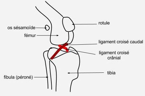
As humans, dog knee joint contains two cruciate ligaments whose role is to ensure the stability of the joint. Each of the two ligaments has a clip on the femur and another on the tibia.
The main function of the cranial cruciate ligament is toprevent the displacement of the tibia to the femur.
Circumstances of occurrence of rupture of the cranial cruciate ligament
In humans the rupture of the cruciate ligaments intervenes, most often, After a major trauma especially during the practice of some sports (Ski, Football, Baseball…)
In dogs, This condition may, in the same way, occur following a"bad move"in a phase of intense gaming or poorly conducted sports activity. However, These acute trauma do not represent the main cause of rupture of the anterior cruciate ligament.Indeed, This failure occurs frequently without having any prior trauma could be noted. It is secondary to a gradual weakening various components of the joint.
Contributing factors
Certain factors predispose to the rupture of the anterior cruciate ligament:
¤ Obesity :more the animal is big, more pressure on his ligaments are important
¤La physical inactivity :very low physical activity and thus a rare request for joint structures makes fragile them ligaments cross
¤ One chronic dislocation of the patella : small breed dogs suffer frequent dislocation of the patella. The latter causes chronic inflammation of the joint and changes forces on ligaments, weakening them.
¤ Finally, theosteoarthritis of the knee is a factor favouring this pathology.
The rupture of the cranial cruciate ligament is accompanied by very frequently concomitant lesions of the menisci.
Highlighting
– The traumatic ruptures cranial cruciate ligament may relate to animals of all ages. The dog then presents a very frank lameness of the Member immediately after the trauma.
– The secondary fractures to a degenerative phenomenon meet, they, in middle-aged animals (6 years, age at which osteoarthritis may already be present) or older. It causes lameness of more gradual onset, not connected to a specific trauma.
During the clinical examination, the veterinarian highlights a pain when it supports on the knee or mobilizes it and may note a swelling of the joint
Cruciate ligament rupture confirmation can be done by several methods :
¤ When handling :
As we stated, the cranial cruciate ligament prevents the tibia forward compared to the femur. When ligament rupture, This "Advanced" becomes possible.
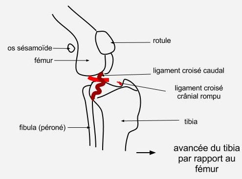
By practising a suitable handling, the veterinarian may highlight this abnormal mobilization of the tibia to the femur. This is called the "sign of the drawer"
This "sign of the drawer" is unfortunately not always present, especially when the inflammation and swelling of the knee limited his handling, When injury associated with a meniscus can skew the review or even when only partial ligament rupture.
¤ By X-ray :
The x-ray will allow suggestive of osteoarthritis and even signs of diagnostiquerune total rupture of the anterior cruciate ligament (ligaments are not directly visible in the picture but the x-ray taken in a suitable position may show an abnormal position of the tibia to the femur). On the other hand, This method does not highlight partial ligament rupture.
¤ Ultrasound :
At the ultrasound, inflammation of the articular membranes can be highlighted and the cranial cruciate ligament visualized. Thus, a suspicion, handling, from rupture of the cranial cruciate ligament will be confirmed and the partial ligament ruptures can be detected.
¤ MRI :
MRI, not only, the display says cruciate ligaments but also the menisci of the knee, What is very interesting knowing that ruptures of the anterior cruciate ligament are very often associated with meniscal lesions.
¤ By Arthroscopy :
Arthroscopic examination performed under general anesthesia. It consists of introducing a miniature camera inside the joint to see all structures. It has several interests:
– It allows to confirm the diagnosis of rupture of the cruciate ligament,
– It allows to visualize the menisci and see if they are injured
– and if the diagnosis is confirmed, the surgeon can treat the rupture of the ligament during anesthesia of Arthroscopy.
Diagnosis and surgical treatment are achievable at the same time.
Treatment
The treatment of rupture of the anterior cruciate ligament is surgical.
The intervention must enable stabilize the joint during walking.
Several methods can be used. The surgeon will decide the most appropriate to each case.
The surgery involves:
¤ To replace the ligament by prosthesis.
The prosthesis can be placed inside or outside the joint and will perform the duties of the ruptured cruciate ligament.
It can be synthetic or biological.
¤ To stabilize the knee joint only in support.
For this, a portion of the tibia will be severed and repositioned using plates and screws in a slightly different direction. These amendments will eliminate the voltage which is usually submitted the cranial cruciate ligament when the animal builds on its hind limb.
The abnormal progress of the tibia at each support is therefore deleted without even needing to replace the ruptured cranial cruciate ligament.
Regardless of the method chosen, one exploration of the Interior of the joint is systematically carried out in order to observe the menisci and withdraw some possibly damaged parts.
Postoperative
Follow-up to surgery, your pet must remain in the rest complete and in a confined space for a period of several weeks to avoid any race, any jump that may prevent proper healing of the joint (the surgeon will tell you precisely these times depending on the surgical approach and departure lesions).
Anti-inflammatory to manage pain.
Walks will be limited, in the premierstemps, In short outputs leash for as the animal makes its needs and the duration of these walks will increase and a recovery gradual and controlled exercise may be undertaken. Control x-rays and examinations will be charged by the surgeon to ensure the normal running of the postoperative.
Furthermore, one monitoring careful the weight the animal will be carried out in order to limit any overweight, which as we have seen, is a factor favouring cruciate ligament ruptures.
Finally, the second knee dogs for which l’osteoarthritis seems to have been the promoting factor ligament rupture can be regularly checked by your vet to ensure that it presents no anomaly.
§
The cranial cruciate ligament rupture is a common pathology including animals too sedentary or overweight. Compliance with good hygiene of life, regular physical activity and a monitored diet can reduce the risk of onset of this condition.
Deal with any persistent lameness your companion, It should be quickly consult your vet Since the rupture of a ligament leads very quickly to important lesions of osteoarthritis.